Neutron Imaging Facility
Neutron Radiography
Neutron radiography is a powerful non-destructive imaging technique for the internal evaluation of materials or components. It involves the attenuation of a neutron beam by an object to be radiographed, and registration of the attenuation process (as an image) digitally or on film.
Neutron radiography is similar in principle to X-ray radiography and is complimentary in the nature of information supplied. The interactions of X-rays and neutrons with matter are fundamentally different, however, forming the basis of many unique applications using neutrons. While X-rays interact with the electron cloud surrounding the nucleus of an atom, neutrons interact with the nucleus itself.
Application of Neutron Radiography
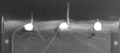
The 0.38 cm thick concrete specimen in the figure below was cut from a 10-cm diameter cylinder that had been loaded to a 3000 micro-cm/cm strain, just beyond its peak stress of 39 MPa. The specimen was impregnated with a Gadolinium Nitrate contrast agent prior to neutron imaging.

Right: Small fractures (microcracks) that were filled with the contrast agent become visible and appear as white lines on the neutron radiograph. Images courtesy of H. Aderhold, Cornell University
NCSU Neutron Imaging Facility (NIF)
The Neutron Imaging Facility is installed at beamport #5 of the PULSTAR reactor, which provides a nominal source flux of 2.5×10¹² n/cm2/sec at 1 MW. The beam is collimated and filtered with 12 inches of single crystal sapphire.
The current aperture is a 1.44”x1.75” (1.8” effective diameter) rectangular cross section opening in a BORAL plate, which yields an L/D ratio of ~140 at the 6.5-meter imaging plane. The resolution of the system is ~ 33 microns for conventional radiographic film. Measurements using ASTM standards show that the NIF achieves a beam quality classification of IA.

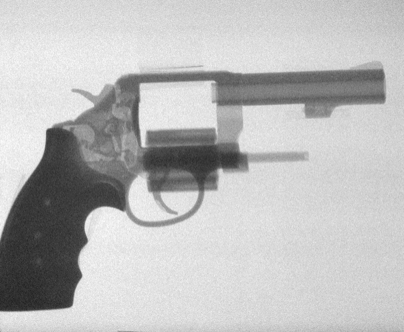
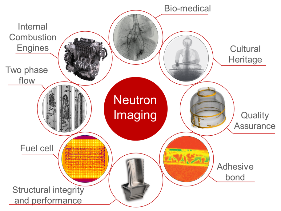
Example Imaging Applications